Hilko WeerdaMedicine

01 A life in medicine

Hilko
Weerda

Physician, surgeon, teacher and scientist.

Read the story
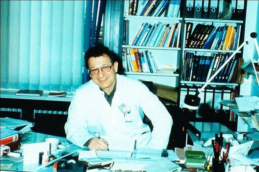
Hilko Weerda seated in his office as Medical Director in Lübeck.
Fig. 96bHilko Weerda in his office as Medical Director of the University ENT Clinic in Lübeck.

His medical work combined artistic creativity with respect for people. Hilko Weerda studied medicine and dentistry and worked as an ENT physician, surgeon, researcher and teacher.

In 1987, he was appointed Medical Director of the University ENT Clinic in Lübeck. He shared his knowledge through courses, books and scientific publications.

His professional life brought together patient care, research, teaching and the development of surgical instruments.

02 Orientation

Eight dates.
One long journey.

Eight milestones in his medical career at a glance.

  1. Degrees in medicine and dentistry
  2. Beginning of ENT specialist training in Freiburg
  3. Habilitation on the cervical trachea
  4. Prototype of the distending laryngoscope
  5. Appointment in Lübeck
  6. International symposium on auricular malformations
  7. Book on plastic-reconstructive surgery
  8. Return to Freiburg

03 Biography · Beginning

From art
to medicine

Before medicine became his central focus, Hilko Weerda studied sculpture with Professor Wimmer and painting with Fritz Griebel at the Nuremberg Academy of Fine Arts in 1958/59. He then began a dual course of study in medicine and dentistry in Erlangen and Munich — initially intending to become a maxillofacial surgeon.

Two doctoral dissertations accompanied this training. He completed both degrees in 1967. Medicine did not replace art; both practices remained part of the same life.

“… Finding fulfilment as an artist in my beloved profession as a physician, scientist and teacher”

Hilko Weerda

Early reconstructive work

In Erlangen, Hilko Weerda worked on facial defect prostheses and made auricular epitheses for children at the university ENT clinic.

The acrylic epitheses available at the time had limitations: the results were not yet satisfactory.

Freiburg and the mentors

In September 1968, he began his ENT specialist training in Freiburg with Professor Fritz Zöllner. From 1970, he worked under Professor Chlodwig Beck.

In Freiburg, he developed his clinical and scientific work further. In 1974, he completed his habilitation on surgery of the cervical trachea.
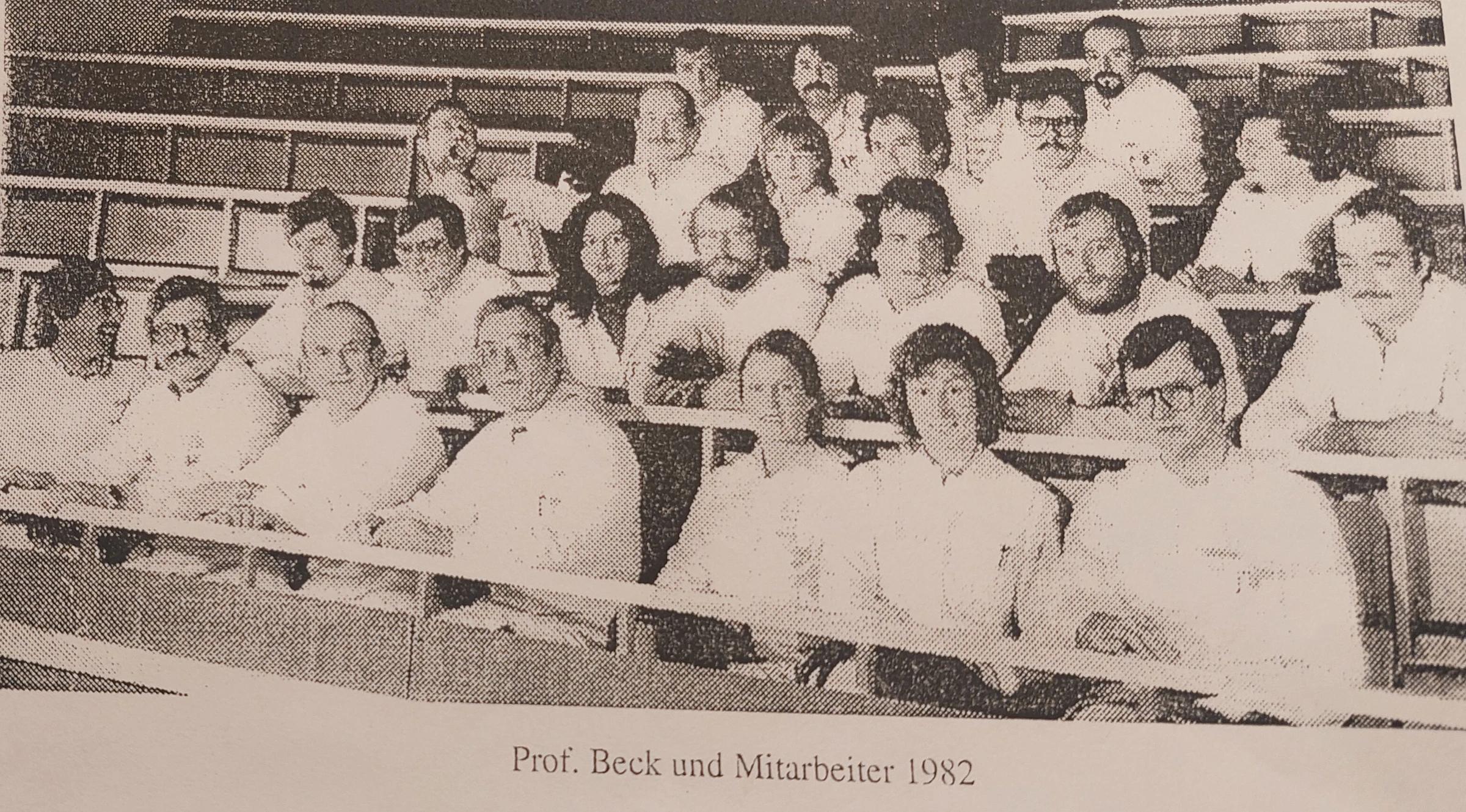
Professor Chlodwig Beck with the Freiburg clinical team in 1982.
Fig. 169bProfessor Chlodwig Beck and the Freiburg team, 1982.Archival transition: from a personal path to the professional environment.

1974 Document

An academic milestone, not an endpoint.

In 1974, Professor Chlodwig Beck presented Hilko Weerda with his habilitation certificate. In Freiburg, Weerda also developed his own aids and instruments for surgical practice.
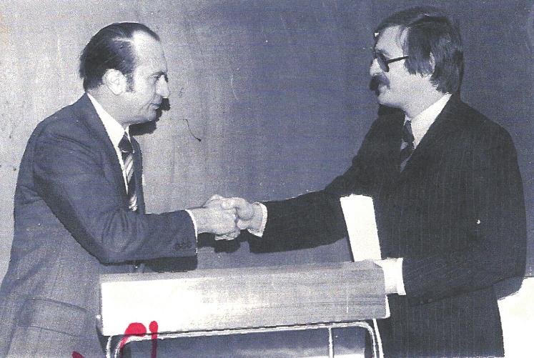
Professor Chlodwig Beck presents Hilko Weerda with his habilitation certificate in 1974.
Fig. 65Professor Chlodwig Beck presents Hilko Weerda with his habilitation certificate, 1974.

04 Instruments & developments

When a clinical task
calls for a form.

In Freiburg, Hilko Weerda developed aids and instruments for specific medical tasks. The first evidence in this chapter traces the path from a workshop model to the later Storz version.

Development path of this instrument

  1. 01 Clinical task
  2. 02 Idea
  3. 03 Prototype
  4. 04 Practical instrument

01 / Prototype

1977: a wider distending laryngoscope

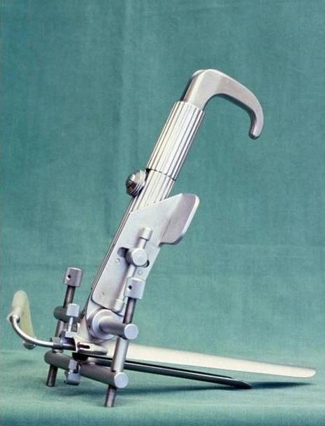
Prototype of the distending laryngoscope, developed by Hilko Weerda with Mr Pedersen in 1977.
Fig. 172aWorkshop prototype, 1977.

For certain endoscopic procedures, the available laryngoscopes were too narrow. Together with workshop manager Mr Pedersen, Hilko Weerda developed this prototype in 1977.

02 / Application

From workshop model to the Storz version

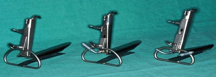
Later version of the Weerda distending laryngoscope developed by Storz for clinical use.
Fig. 172bLater Storz version.

The instrument manufacturer Storz further developed the model for clinical use. The two archival images make the transition from an individual workshop model to the later instrument series immediately understandable.

Prototype of the distending laryngoscope, developed by Hilko Weerda with Mr Pedersen in 1977.
Fig. 172aWorkshop prototype · 1977
Later version of the Weerda distending laryngoscope developed by Storz for clinical use.
Fig. 172bLater Storz version

Prototype

practical instrument

04 Further instruments & developments

Not one line.
Multiple tasks.

The following developments arose from different clinical situations. What they share is not a single method, but a precise response to a practical problem.

A

Airway · Trachea

Support, open,
create access.

From small support rings to instruments for tracheotomy, this section places material, access and handling side by side — not as a product family, but as responses to different tasks.

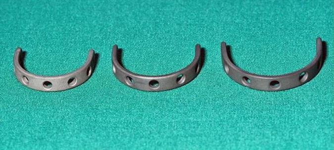
Three curved titanium support rings for the trachea.
Fig. 170b

Titanium support rings as a later, more stable design for the trachea.

Fig. 170c

Technical documentation of the position and application of the tracheal splints.

02 / Material & system

From an individual component to a supporting system.

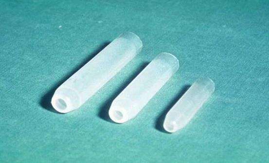
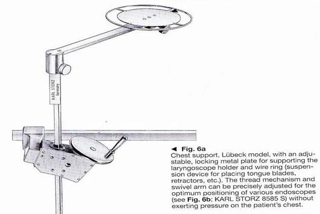
Experimental silicone tubes were intended to support single-stage reconstruction of the trachea. The “Lübeck” chest support was developed for endoscopy and suspended instruments.

Three experimental silicone tubes in different sizes.
Fig. 171a

Experimental silicone tubes.

Technical drawing of the Lübeck chest support for endoscopic instruments.
Fig. 173a

“Lübeck” chest support as a system drawing.

03 / Instrument & application

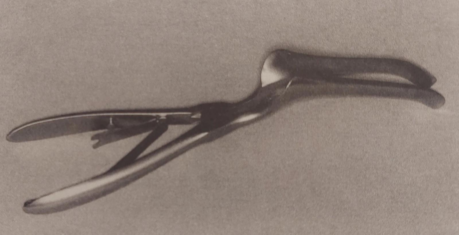
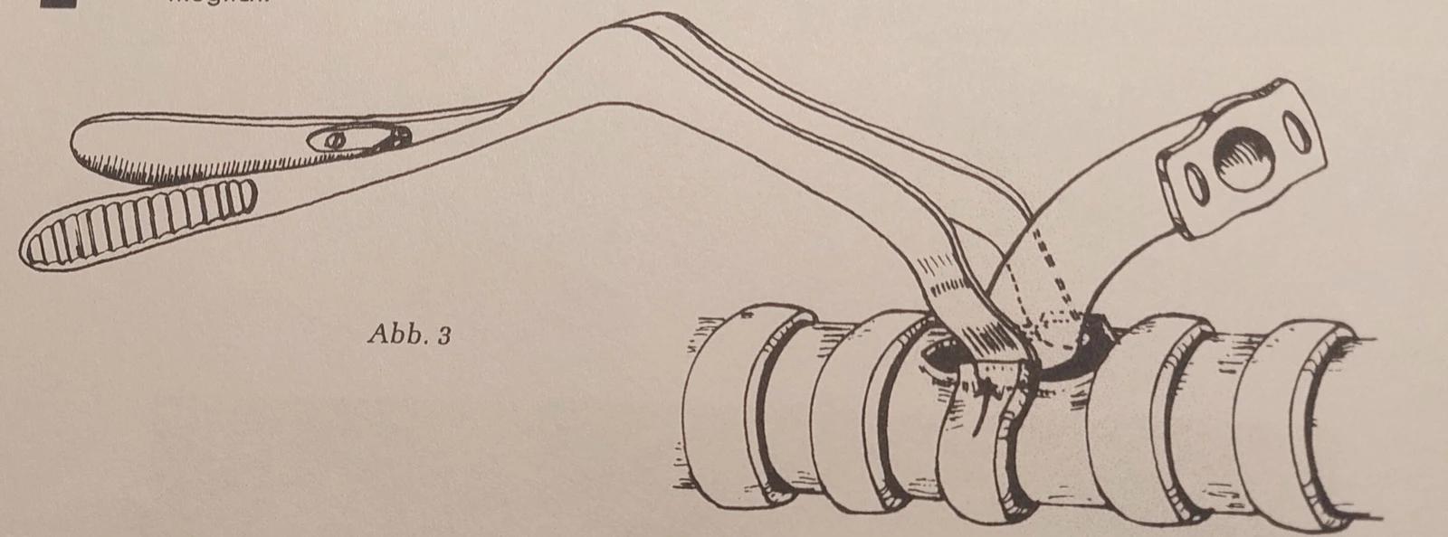
Spreading speculum

Developed to facilitate the insertion of a tracheal cannula during tracheotomy.

Spreading speculum for tracheotomy.
Fig. 174a

The instrument.

Drawing showing the application of the spreading speculum to the trachea.
Fig. 174b

The application shown in a drawing.

04 / Instrument & application

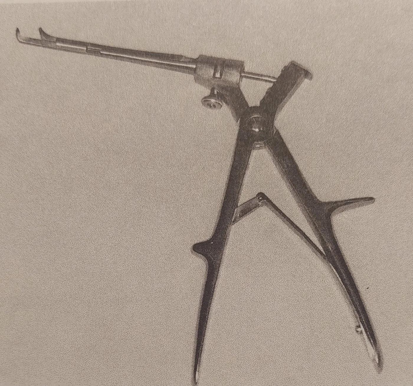
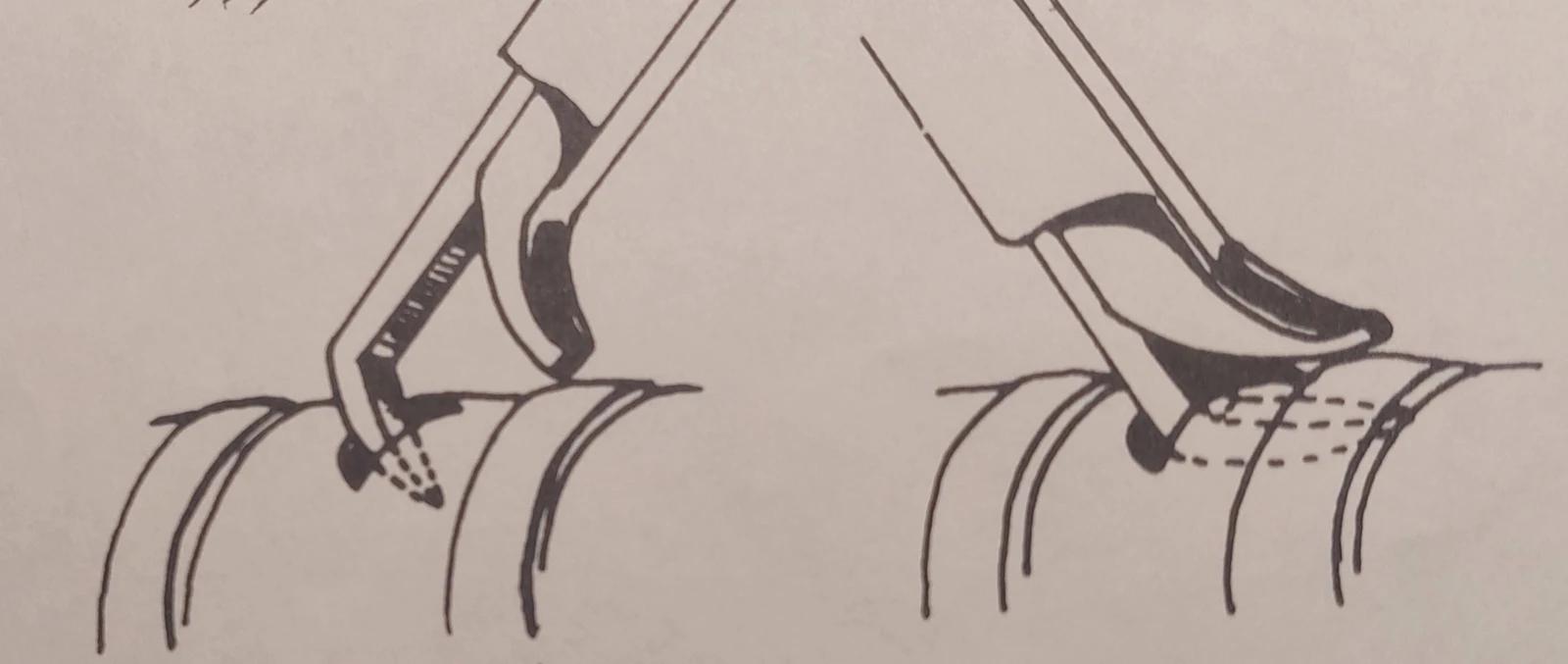
Tracheotomy punch

A specialised instrument with its own application documented in a drawing.

Special punch for tracheotomy.
Fig. 175a

Tracheotomy punch.

Drawing showing the use of the tracheotomy punch.
Fig. 175b

Documented application step.

B

Precision · Detail

Tools for
fine procedures.

Dermatome, forceps and scissors appear as a related study group — closer to a collection of precise solutions than a product overview.

C

Instrument · Documentation · System

When a tool
leads to further work.

Two relationships conclude the technical chapter: a diverticuloscope leads to documentation; an especially thin tube calls for a supporting ventilation system.

Story 01

Diverticuloscope → Documentation

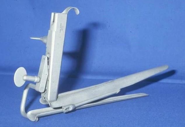
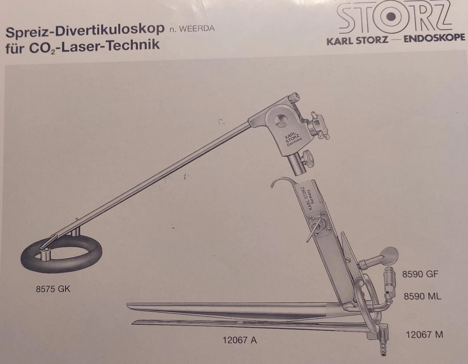
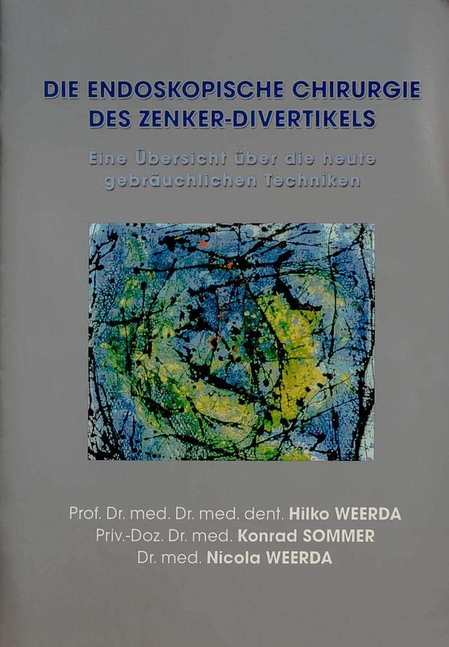
The Weerda distending diverticuloscope does not stand alone here. A technical illustration and the later publication with Nicola Weerda and Conrad Sommer show how the instrument also became communicable knowledge.

Weerda distending diverticuloscope on a blue background.
Fig. 179a

Distending diverticuloscope.

Technical Storz illustration of the Weerda distending diverticuloscope.
Fig. 179b

Technical illustration by Storz.

Cover of the publication Die endoskopische Chirurgie des Zenker-Divertikels.
Fig. 180

Hilko Weerda, Conrad Sommer and Nicola Weerda: publication on endoscopic diverticular surgery.

Story 02

3 mm tube → Ventilator

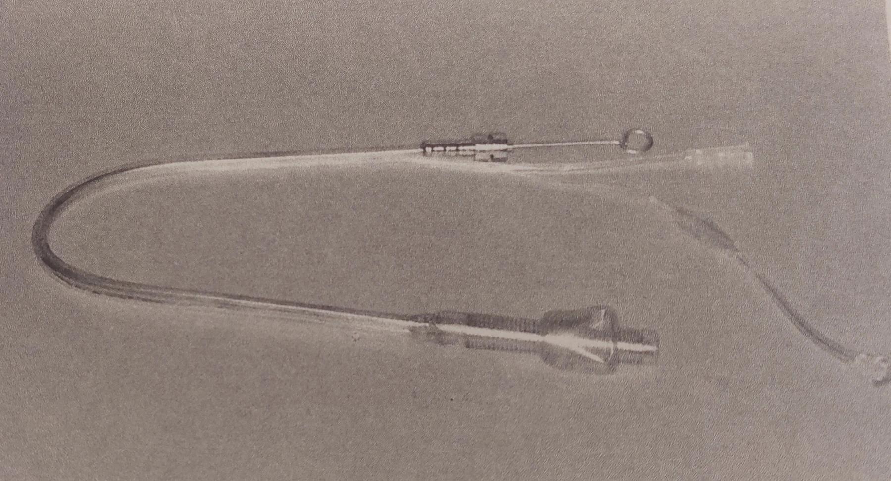
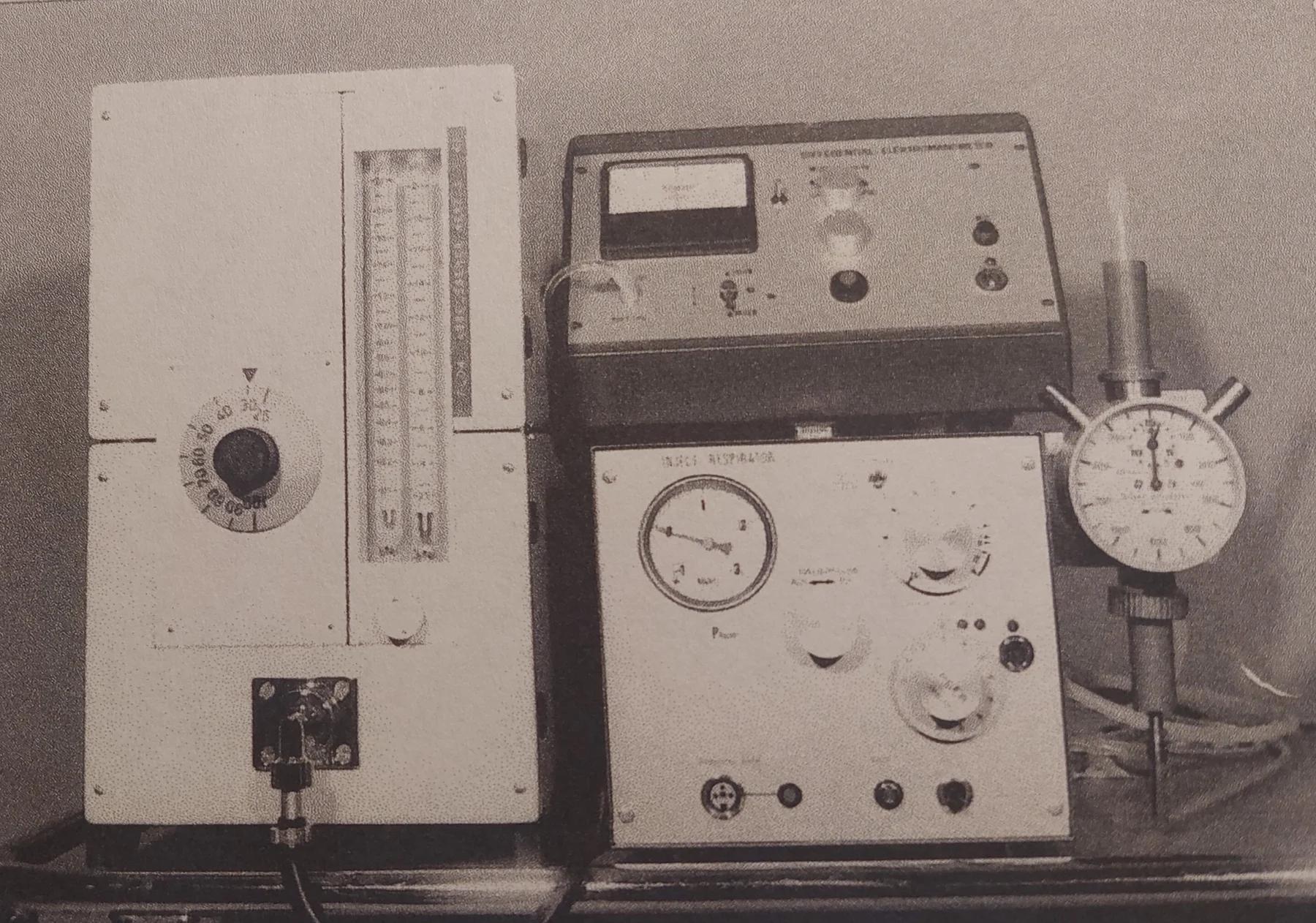
A thin intubation tube was developed to provide an unobstructed view during endoscopic procedures. A ventilator was developed for it together with Mr Pedersen. The two images form a single system.

Three-millimetre intubation tube for injection ventilation.
Fig. 181

3 mm intubation tube for injection ventilation.

Ventilator developed with Mr Pedersen for thin intubation tubes.
Fig. 182

Ventilator developed with Mr Pedersen.

Task

Tool

Transmission

05 Courses · Teaching · Network

Knowledge becomes
a shared practice.

Through courses in Lübeck and Munich and the 1997 symposium, Hilko Weerda shared surgical experience and encouraged professional exchange. The invited international specialists included Burt Brent, Satoru Nagata and Françoise Firmin, among others.

Teaching in everyday practice

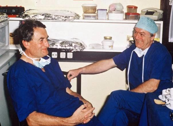
In the archive, the courses appear as more than programmes or teaching texts. An informal moment with Otto Staindl and Ernst Kastenbauer reveals the collegial setting behind the transfer of specialist knowledge.

Otto Staindl and Ernst Kastenbauer during a course in Lübeck.
Fig. 185

Otto Staindl and Ernst Kastenbauer during a course in Lübeck.

06 Books & publications

Courses become
books.

Manuscripts, surgical drawings and clinical experience developed into compendia, specialist books and translations. This selection traces an evolution — it is not a complete library.

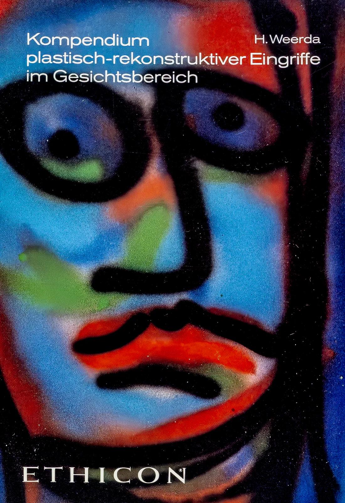
Cover of the Kompendium plastisch-rekonstruktiver Eingriffe im Gesichtsbereich.
Fig. 183

Kompendium plastisch-rekonstruktiver Eingriffe im Gesichtsbereich.

From course manuscript to compendium

Draw, organise, pass on.

The manuscript for his training courses became a compendium containing Hilko Weerda’s own surgical drawings. The first edition appeared in 1984; four editions were published in total.

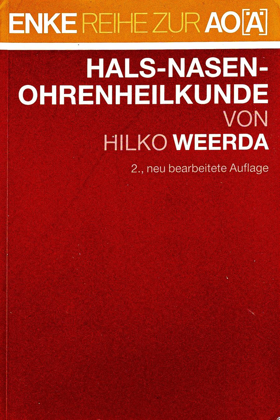
Cover of Hilko Weerda’s specialist book Hals-Nasen-Ohrenheilkunde.
Selection 01

Hals-Nasen-Ohrenheilkunde.

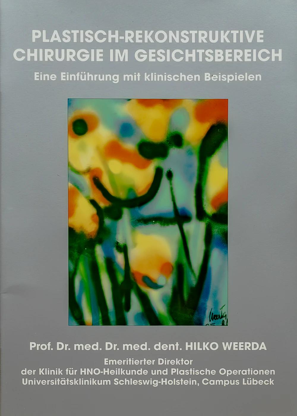
Cover of the publication Plastisch-Rekonstruktive Chirurgie im Gesichtsbereich.
Selection 02

Plastisch-Rekonstruktive Chirurgie im Gesichtsbereich.

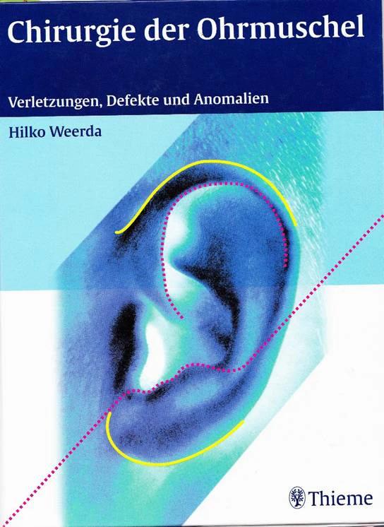
German cover of the book Chirurgie der Ohrmuschel.
Selection 03

Chirurgie der Ohrmuschel.

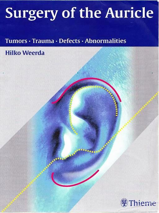
English cover of the book Surgery of the Auricle.
Selection 04

Surgery of the Auricle.

Scientific work

Hilko Weerda published more than 300 publications during his years in Freiburg and Lübeck.

The selected books represent this scientific work; they do not constitute a complete bibliography.

07 Recognition & scientific legacy

What remains
continues.

Hilko Weerda’s work as a surgeon, researcher and teacher earned professional recognition over many years. His instruments, publications and training courses helped pass on surgical knowledge.

Honours & memberships

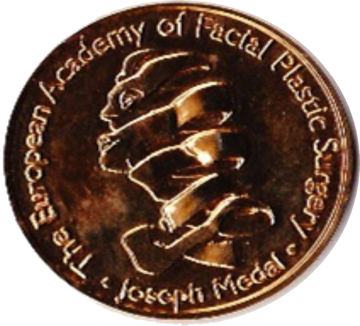
Jacques Joseph Medal

Hilko Weerda is a co-founder of the European Academy of Facial Plastic Surgery, an honorary member since 2008 and a recipient of the Jacques Joseph Medal.

He also became an honorary member of the German Society of Otorhinolaryngology and, in 2018, honorary president during Professor Wollenberg’s presidency.

Bronze-coloured Jacques Joseph Medal with a plastic-surgery profile motif.
Fig. 188

Jacques Joseph Medal.

2002 Epilogue

Hilko and Barbara Weerda returned to Freiburg. After his return, he assisted his daughter-in-law Nicola, who had also chosen to become an ENT physician, with difficult auricular reconstructions — until around the age of eighty.

After his return to Freiburg, art once again became a greater part of daily life: the studio, exhibitions and engagement with the collection. Medicine and art appear as two forms of his creative work within a single life.

To Art